آخر المواضيع المضافة

 النبات
الحيوان
الأحياء المجهرية
علم الأمراض
التقانة الإحيائية
التقنية الحيوية المكروبية
التقنية الحياتية النانوية
علم الأجنة
الأحياء الجزيئي
علم وظائف الأعضاء
الغدد
المضادات الحيوية
النبات
الحيوان
الأحياء المجهرية
علم الأمراض
التقانة الإحيائية
التقنية الحيوية المكروبية
التقنية الحياتية النانوية
علم الأجنة
الأحياء الجزيئي
علم وظائف الأعضاء
الغدد
المضادات الحيوية| Flow cytometry |


|
|
Read More
Date: 28-10-2015
Date: 28-12-2020
Date: 1-11-2015
|
Flow cytometry is a technique used to measure the chemical and physical characteristics of single cells in solution. It is used in different fields such as molecular biology, immunology, virology, parasitology, and microbiology, in research and clinical settings. In flow cytometry, a cell suspension is injected into the central channel of a fluid system that is surrounded by a physiological buffer of fast-flowing fluid known as the sheath fluid (Suthanthiraraj & Graves, 2013).
As each cell goes through a laser beam, physical structures present in the cell may be struck, resulting in the scattering of light (Cossarizza & Chang, 2018). Forward scatter (FS) is measured by a detector in front of the light beam (McKinnon, 2018; Mealey & Long, 2018). Side scatter (SS) is measured by detectors found on the side of the light beam. FS is proportional to the size of the cell or cell surface (McKinnon, 2018; Mealey & Long, 2018). SS is proportional to cell granularity or internal complexity (Reggeti & Bienzle, 2011). FS and SS can be used to differentiate different cell types in a heterogeneous cell population. Although the light scatter signal is influenced by particle size, other factors such as the wavelength of the laser, refractive index, and sheath fluid can contribute to the light scatter signal (Kerker, 1983; Loken & Houck, 1981; Reardon et al., 2014; Salzman et al., 1979). The addition of fluorescent dyes, for example, propidium iodide or fluorochromes (e.g., fluorescein isothiocyanate, phycoerythrin) to cells can give quantitative and qualitative information relating to the structural proper ties of cells (Cossarizza & Chang, 2018; McKinnon, 2018). Fluorochromes are commonly conjugated to antibodies recognizing specific cell surface markers. In fluorescence flow cytometry, a peak is produced as the fluorescent labeled cells pass through the laser beam (Adan et al., 2017). Each fluorescent dye or fluorochrome has its characteristic excitation and emission spectra. Every cell in a sample that passes through the laser intercept is recorded as a distinct event (McKinnon, 2018). The intensity or amount of scattered light and fluorescent emissions is detected by photodiodes or photo multiplier tubes and converted into a voltage pulse (McKinnon, 2018). Voltage pulses are then converted to channel numbers and stored by the cytometer computer system for analysis by a flow cytometry analysis program (McKinnon, 2018; Mealey & Long, 2018).
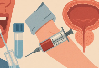
Flow cytometry can be used to detect viral proteins such as the SARS CoV-2 spike protein. (Lapuente et al. 2020) used flow cytometry to detect IgG and IgM antibodies against SARS-CoV-2 spike proteins in human serum samples (Lapuente et al., 2020). Antibodies against SARS-CoV-2 present in the serum of patients bound to human embryonic kidney cells expressing the SARS-CoV-2 spike protein (Lapuente et al., 2020). Bound antibodies were then incubated with secondary detection antibodies and analyzed using flow cytometry (Lapuente et al., 2020). Results showed that the flow cytometry assay detected 100% of IgM and IgG positive samples, with similar sensitivity to commercial ELISA and chemiluminescent immunoassays kits (Lapuente et al., 2020). The flow cytometry assay was also highly sensitive in detecting antibodies in PCR-confirmed SARS-CoV-2 positive patients (Lapuente et al., 2020). Thus the flow cytometry based serological assay developed by Lapuente et al. (2020) can be used in seroepidemiological studies and evaluation of immunity of patients after vaccination.
General flow cytometry protocol
A detailed general protocol on flow cytometry (Fig. 1) can be found below. This protocol was adapted from Biorad, and the Yale flow cytometry proto col (Biorad, 2016; Yale, 2019). Samples are injected in sheath fluid, which focuses samples to the point of laser intercept. Fluorescence signals and forward and side scattered light are recorded for each cell as it passes through the laser beam.
Materials
Cells to be stained, PBS/BSA (containing 1% BSA), Fc blocking solution, antibodies (primary and secondary antibody), Fc block, and 5 mL falcon tubes.
Flow cytometry procedure
1. Harvest, wash cells, and adjust cell number to a concentration between 13105 to 13107 cells/mL in ice-cold PBS/BSA.
2. Place 100 μL of cell suspension in a tube.
3. Add primary antibody to the cell suspension and incubate for 30 minutes.
4. Wash cells with PBS/BSA three times.
5. Centrifuge at 300 -400 g for 5 minutes at room temperature and discard the supernatant.
6. Add labeled secondary antibody and incubate in the dark for approximately 1 hour.
7. Wash cells with PBS/BSA three times.
8. Centrifuge at 300- 400 g for 5 minutes at room temperature and discard the supernatant.
9. Include control tubes in the experiment, one tube should contain only the secondary antibody and the other tube should not contain an antibody.
10. Resuspend cells in 200 μL to 1 mL of cold (4C) PBS or with 200 μL of 0.5 mL paraformaldehyde in PBS if needed.
11. Keep samples on ice and run samples on the flow cytometer.
In flow cytometry, unstained cells should be included in the experiment to check for autofluorescence. Fc block should be added to cells that express high levels of Fc receptors to avoid nonspecific binding. This is a general protocol and hence cell preparations, controls, and antibody dilutions will need to be evaluated before the experiment.

Fig1. Schematic representation of flow cytometry.
References
-------------
Adan, A., Alizada, G., Kiraz, Y., Baran, Y., & Nalbant, A. (2017). Flow cytometry: Basic princi ples and applications. Critical Reviews in Biotechnology, 37(2), 163 176. Available from https://doi.org/10.3109/07388551.2015.1128876.
Biorad. (2016). Indirect immunofluorescence staining of cells & blood protocol | Bio-Rad. Available from https://www.bio-rad-antibodies.com/indirect-immunofluorescence-staining of-cells-blood-for-flow-cytometry.html.
Cossarizza, A., & Chang, H. (2018). Guidelines for the use of flow cytometry and cell sorting in immunological studies (second edition). Physiology & Behavior, 176(1), 139 148. Available from https://doi.org/10.1117/12.2549369.Hyperspectral.
Kerker, M. (1983). Elastic and inelastic light scattering in flow cytometry. Cytometry, 4(1), 1 10. Available from https://doi.org/10.1002/cyto.990040102.
Lapuente, D., Maier, C., Irrgang, P., Hu ¨bner, J., Peter, A. S., Hoffmann, M., Ensser, A., Ziegler, K., Winkler, T. H., Birkholz, T., Kremer, A. E., Steininger, P., Korn, K., Neipel, F., U ¨berla, K., & Tenbusch, M. (2020). Rapid response flow cytometric assay for the detection of antibody responses to SARS-CoV-2. European Journal of Clinical Microbiology and Infectious Diseases,1 9. Available from https://doi.org/10.1007/s10096-020-04072-7.
Loken, M. R., & Houck, D. W. (1981). Light scattered at two wavelengths can discriminate via ble lymphoid cell populations on a fluorescence-activated cell sorter. Journal of Histochemistry & Cytochemistry, 29(5), 609 615. Available from https://doi.org/10.1177/ 29.5.7252128.
McKinnon, K. M. (2018). Flow cytometry: An overview. Current Protocols in Immunology, 2018. Available from https://doi.org/10.1002/cpim.40, 5.1.1-5.1.11.
Reardon, A. J. F., Elliott, J. A. W., & McGann, L. E. (2014). Fluorescence as an alternative to light-scatter gating strategies to identify frozen-thawed cells with flow cytometry. Cryobiology, 69(1), 91 99. Available from https://doi.org/10.1016/j.cryobiol.2014.05.009.
Reggeti, F., & Bienzle, D. (2011). Flow cytometry in veterinary oncology. Veterinary Pathology, 48, 223 235. Available from https://doi.org/10.1177/0300985810379435.
Salzman, G. C., Wilder, M. E., & Jett, J. H. (1979). Light scattering with stream-in-air flow systems. Journal of Histochemistry & Cytochemistry, 27(1), 264 267. Available from https:// doi.org/10.1177/27.1.374583.
Suthanthiraraj, P., & Graves, S. (2013). Fluidics. Current protocols in cytometry, 01(Suppl. 65), Unit-Unit. Available from https://doi.org/10.1002/0471142956.cy0102s65.


|
|
|
|
التوتر والسرطان.. علماء يحذرون من "صلة خطيرة"
|
|
|
|
|
|
|
مرآة السيارة: مدى دقة عكسها للصورة الصحيحة
|
|
|
|
|
|
|
نحو شراكة وطنية متكاملة.. الأمين العام للعتبة الحسينية يبحث مع وكيل وزارة الخارجية آفاق التعاون المؤسسي
|
|
|