Heme degradation
After ~120 days in the circulation, RBC are taken up and degraded by the mononuclear phagocyte system (MPS), particularly in the liver and spleen (Fig. 1). Approximately 85% of heme destined for degradation comes from senescent RBC. The remainder is from the degradation of hemeproteins other than Hb.

Figure 1: Formation of bilirubin from heme and its conversion to bilirubin diglucuronide. UDP = uridine diphosphate; Fe = iron; CO = carbon monoxide; NADP(H) = nicotinamide adenine dinucleotide phosphate.
1. Bilirubin formation: The first step in the degradation of heme is catalyzed by microsomal heme oxygenase in macrophages of the MPS.
In the presence of nicotinamide adenine dinucleotide phosphate and oxygen, the enzyme catalyzes three successive oxygenations that result in opening of the porphyrin ring (converting cyclic heme to linear biliverdin), production of carbon monoxide (CO), and release of Fe2+ (see Fig. 1). [Note: The CO has biologic function, acting as a signaling molecule and anti-inflammatory. Biliverdin, a green pigment, is reduced, forming the red-orange bilirubin. Bilirubin and its derivatives are collectively termed bile pigments. [Note: The changing colors of a bruise reflect the varying pattern of intermediates that occurs during heme degradation.]
Bilirubin, unique to mammals, appears to function at low levels as an antioxidant. In this role, it is oxidized to biliverdin, which is then reduced by biliverdin reductase, regenerating bilirubin.
2. Bilirubin uptake by the liver: Because bilirubin is only slightly soluble in plasma, it is transported through blood to the liver by binding noncovalently to albumin. [Note: Certain anionic drugs, such as salicylates and sulfonamides, can displace bilirubin from albumin, permitting bilirubin to enter the central nervous system (CNS). This causes the potential for neural damage in infants .] Bilirubin dissociates from the carrier albumin molecule, enters a hepatocyte via facilitated diffusion, and binds to intracellular proteins, particularly the protein ligandin.
3. Bilirubin diglucuronide formation: In the hepatocyte, bilirubin solubility is increased by the sequential addition of two molecules of glucuronic acid in a process called conjugation. The reactions are catalyzed by microsomal bilirubin UDP-glucuronosyltransferase (bilirubin UGT) using uridine diphosphate (UDP)-glucuronic acid as the glucuronate donor. The bilirubin diglucuronide product is referred to as conjugated bilirubin (CB). [Note: Varying degrees of deficiency of bilirubin UGT result in Crigler-Najjar I and II and Gilbert syndrome, with Crigler-Najjar I being the most severe.]
4. Bilirubin secretion into bile: CB is actively transported against a concentration gradient into the bile canaliculi and then into the bile. This energy-dependent, rate-limiting step is susceptible to impairment in liver disease. [Note: A rare deficiency in the protein required for transport of CB out of the liver results in Dubin-Johnson syndrome.] Unconjugated bilirubin (UCB) is normally not secreted into bile.
5. Urobilin formation in the intestine: CB is hydrolyzed and reduced by gut bacteria to yield urobilinogen, a colorless compound. Most of the urobilinogen is further oxidized by bacteria to stercobilin, which gives feces the characteristic brown color. However, some is reabsorbed from the gut and enters the portal blood. A portion of this urobilinogen participates in the enterohepatic urobilinogen cycle in which it is taken up by the liver and then resecreted into the bile. The remainder of the urobilinogen is transported by the blood to the kidney, where it is converted to yellow urobilin and excreted, giving urine its characteristic color. The metabolism of bilirubin is summarized in Figure 2.
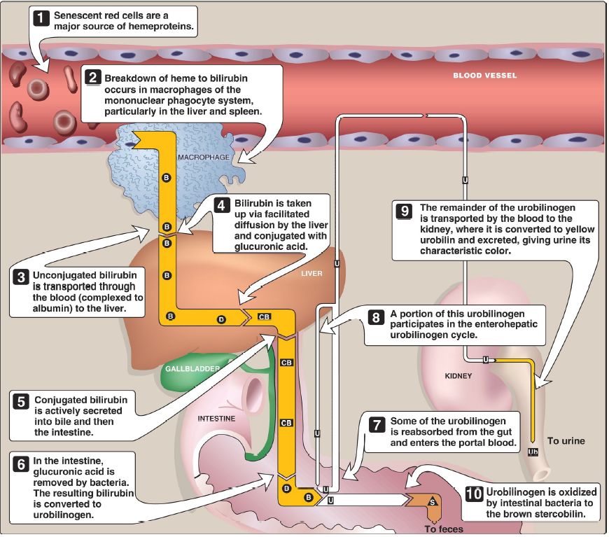
Figure 2: Catabolism of heme. = bilirubin; = conjugated bilirubin; = urobilinogen; = urobilin; = stercobilin.


