آخر المواضيع المضافة

 النبات
الحيوان
الأحياء المجهرية
علم الأمراض
التقانة الإحيائية
التقنية الحيوية المكروبية
التقنية الحياتية النانوية
علم الأجنة
الأحياء الجزيئي
علم وظائف الأعضاء
الغدد
المضادات الحيوية
النبات
الحيوان
الأحياء المجهرية
علم الأمراض
التقانة الإحيائية
التقنية الحيوية المكروبية
التقنية الحياتية النانوية
علم الأجنة
الأحياء الجزيئي
علم وظائف الأعضاء
الغدد
المضادات الحيوية| Angle of the Eye-Iridocorneal Angle |


|
|
Read More
Date: 27-7-2016
Date: 19-1-2017
Date: 5-1-2017
|
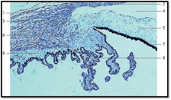
Angle of the Eye-Iridocorneal Angle
The upper part of the figure shows the substantiae propriae sclerae 1 and the cornea 2 . The slits in the sclera represent the canal of Schlemm (sinus venosus sclerae). The ciliary body with the ciliary muscle 6 forms lamella-shape d ciliary processes with a thin pars plana and a pars plicata. The processes are covered by a two-layered epithelium, which is thought to produce the intraocular fluid. The pars plicata 8 features about 70 ciliary processes .
1 Substantia propria sclerae
2 Substantia propria corneae
3 Angle of the eye (Schwalbe’s line)
4 Anterior chamber of the eye
5 Iris
6 Ciliary muscle
7 Pigmented epithelium of the iris
8 Ciliary processus
Stain: hematoxylin-eosin; magnification: × 40
References
Kuehnel, W.(2003). Color Atlas of Cytology, Histology, and Microscopic Anatomy. 4th edition . Institute of Anatomy Universitätzu Luebeck Luebeck, Germany . Thieme Stuttgart · New York .


|
|
|
|
التوتر والسرطان.. علماء يحذرون من "صلة خطيرة"
|
|
|
|
|
|
|
مرآة السيارة: مدى دقة عكسها للصورة الصحيحة
|
|
|
|
|
|
|
نحو شراكة وطنية متكاملة.. الأمين العام للعتبة الحسينية يبحث مع وكيل وزارة الخارجية آفاق التعاون المؤسسي
|
|
|