آخر المواضيع المضافة

 النبات
الحيوان
الأحياء المجهرية
علم الأمراض
التقانة الإحيائية
التقنية الحيوية المكروبية
التقنية الحياتية النانوية
علم الأجنة
الأحياء الجزيئي
علم وظائف الأعضاء
الغدد
المضادات الحيوية
النبات
الحيوان
الأحياء المجهرية
علم الأمراض
التقانة الإحيائية
التقنية الحيوية المكروبية
التقنية الحياتية النانوية
علم الأجنة
الأحياء الجزيئي
علم وظائف الأعضاء
الغدد
المضادات الحيوية| Th2 Cells |


|
|
Read More
Date: 2025-03-25
Date: 1-11-2015
Date: 8-12-2020
|
Th2 cells are induced by parasitic worm infections and promote IgE-, mast cell- and eosinophil- mediated destruction of these parasites (Fig. 1). The signature cytokines of Th2 cells—IL-4, IL-5, and IL-13—function cooperatively in eradicating worm infections. Helminths are too large to be phagocytosed, so mechanisms other than macrophage activation are needed for their destruction. When Th2 and related Tfh cells encounter the antigens of helminths, the T cells secrete their cytokines. IL-4 produced by Tfh cells stimulates the production of IgE antibodies, which coat the helminths and thus help in their clearance. Eosinophils use their Fc receptors to bind to the IgE and are activated by IL-5 produced by the Th2 cells, as well as by signals from these IgE-specific Fc receptors. Activated eosinophils release their granule con tents, which are toxic to the parasites. IL-13 stimulates mucus secretion and intestinal peristalsis, increasing the expulsion of parasites from the intestines. IgE also binds to mast cells and is responsible for their activation, leading to the secretion of chemical mediators that stimulate inflammation and proteases that destroy toxins.

Fig1. Functions of Th2 cells. Th2 cells produce the cytokines interleukin-4 (IL-4), IL-5, and IL-13. IL-4 (and IL-13) act on B cells to stimulate production mainly of IgE antibodies, which bind to mast cells. Help for antibody production may be provided by Tfh cells that produce Th2 cytokines and reside in lymphoid organs and not by classical Th2 cells. IL-5 activates eosinophils, a response that is important in the destruction of helminths. APC, Antigen-presenting cell; Ig, immunoglobulin; IL, interleukin.
T h2 cytokines inhibit classical macrophage activation and stimulate the alternative pathway of macro phage activation (Fig. 2). IL-4 and IL-13 shut down the activation of inflammatory macrophages, thus terminating these potentially damaging reactions. These cytokines also can activate macrophages to secrete growth factors that act on fibroblasts to increase collagen synthesis and induce fibrosis. This type of macrophage response is called alternative macrophage activation, to distinguish it from classical activation, which enhances microbicidal functions. Alternative macrophage activation mediated by Th2 cytokines may play a role in tissue repair following injury and may con tribute to fibrosis in a variety of disease states.

Fig2. Classical and alternative macrophage activation. Classically activated (M1) macrophages are induced by microbial products binding to TLRs and cytokines, particularly interferon-γ (IFN-γ), and are microbicidal and proinflammatory. Alternatively activated (M2) macrophages are induced by interleukin-4 (IL-4) and IL-13 (produced by certain subsets of T lymphocytes and other leukocytes) and are important in tissue repair and fibrosis. The M1 and M2 populations may represent extreme phenotypes, and there may be other macro phage populations that express different sets of proteins. Also, in most immune responses, various mixtures of these macrophages are likely induced. NO, Nitric oxide; ROS, reactive oxygen species; TGF-β, transforming growth factor β; TLR, Toll-like receptor.
Th2 cells are involved in allergic reactions to environmental antigens. The antigens that elicit such reactions are called allergens. They induce Th2 responses in genetically susceptible individuals, and repeat exposure to the allergens triggers mast cell and eosinophil activation. Allergies are the most common type of immune disorder. Antagonists of IL-5 are approved for the treatment of asthma, and an antibody against the IL-4 receptor is approved for the allergic disease atopic dermatitis.
The relative activation of Th1 and Th2 cells in response to an infectious microbe may determine the outcome of the infection (Fig. 3). For example, the protozoan parasite Leishmania major lives inside the phagocytic vesicles of macrophages, and its elimination requires the activation of the macrophages by L. major specific Th1 cells. Most inbred strains of mice make an effective Th1 response to the parasite and are thus able to eradicate the infection. However, in some inbred mouse strains, the response to L. major is dominated by Th2 cells, and these mice succumb to the infection. Mycobacterium leprae, the bacterium that causes leprosy, is a pathogen for humans that also lives inside macrophages and may be eliminated by cell-mediated immune mechanisms. Some people infected with M. leprae are unable to eradicate the infection, which, if left untreated, will progress to a destructive form of the disease, called lepromatous leprosy. By contrast, in other patients, the bacteria induce strong cell-mediated immune responses with activated T cells and macrophages around the infection site and few surviving microbes; this form of less injurious infection is called tuberculoid leprosy.

Fig3. Balance between Th1 and Th2 cell activation determines outcome of intracellular infections. Naive CD4+ T lymphocytes may differentiate into Th1 cells, which activate phagocytes to kill ingested microbes, and Th2 cells, which inhibit classical macrophage activation. The balance between these two sub sets may influence the outcome of infections, as illustrated by Leishmania infection in mice and leprosy in humans. IFN, Interferon; IL, interleukin; TNF, tumor necrosis factor.
The tuberculoid form is associated with the activation of M. leprae–specific Th1 cells, whereas the destructive lepromatous form is associated with a defect in Th1 cell activation and sometimes a strong Th2 response. The same principle—that the T cell cytokine response to an infectious pathogen is an important determinant of the outcome of the infection—may be true for other infectious diseases.
Development of Th2 Cells Differentiation of naive CD4+ T cells to Th2 cells is stimulated by IL-4, which may be produced by mast cells, other tissue cells, and T cells themselves at sites of helminth infection ( Fig. 4). IL-4 activates the transcription factor Stat6 and antigen-induced signals in combination with IL-4 induce expression of a transcription factor GATA-3, which is required for Th2 differentiation. Analogous to Th1 cells, these transcription factors stimulate the expression of Th2 cytokines and proteins involved in cell migration and thus promote Th2 responses. IL-4 produced by Th2 cells enhances further Th2 differentiation, thus amplifying the Th2 response.

Fig4. Dendritic cells and other immune cells that respond to different types of microbes secrete cytokines that induce the development of antigen-activated CD4+ T cells into Th2.


|
|
|
|
التوتر والسرطان.. علماء يحذرون من "صلة خطيرة"
|
|
|
|
|
|
|
مرآة السيارة: مدى دقة عكسها للصورة الصحيحة
|
|
|
|
|
|
|
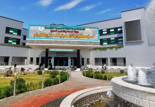
نحو شراكة وطنية متكاملة.. الأمين العام للعتبة الحسينية يبحث مع وكيل وزارة الخارجية آفاق التعاون المؤسسي
|
|
|