آخر المواضيع المضافة

 النبات
الحيوان
الأحياء المجهرية
علم الأمراض
التقانة الإحيائية
التقنية الحيوية المكروبية
التقنية الحياتية النانوية
علم الأجنة
الأحياء الجزيئي
علم وظائف الأعضاء
الغدد
المضادات الحيوية
النبات
الحيوان
الأحياء المجهرية
علم الأمراض
التقانة الإحيائية
التقنية الحيوية المكروبية
التقنية الحياتية النانوية
علم الأجنة
الأحياء الجزيئي
علم وظائف الأعضاء
الغدد
المضادات الحيوية| Th1 Cells |


|
|
Read More
Date: 2025-03-10
Date: 2025-01-27
Date: 8-12-2020
|
The Th1 subset is induced by microbes that are ingested by and activate phagocytes, primarily macro phages, and Th1 cells stimulate phagocyte-mediated killing of ingested microbes (Fig. 1). The signature cytokine of Th1 cells is interferon-γ (IFN-γ), the most potent macrophage-activating cytokine known. (Despite its similar name, IFN-γ is a much less potent antiviral cytokine than the type I IFNs .

Fig1. Functions of Th1 cells. Th1 cells produce the cytokine interferon-γ (IFN-γ), which activates macrophages to kill phagocytosed microbes (classical pathway of macrophage activation). In mice, IFN-γ stimulates the production of IgG antibodies, but this has not been established in humans. APC, Antigen-presenting cell.
Th1 cells, acting through CD40 ligand and IFN-γ, increase the ability of macrophages to kill phagocytosed microbes (Fig. 2). Macrophages ingest and attempt to destroy microbes as part of the innate immune response. The efficiency of this process is greatly enhanced by the interaction of Th1 cells with the macrophages. When microbes are ingested into phagosomes of the macrophages, microbial peptides are presented on class II MHC molecules and are recognized by CD4+ T cells. If these T cells belong to the Th1 subset, they are induced to express CD40 ligand (CD40L, or CD154) and to secrete IFN-γ. Binding of CD40L to CD40 on macrophages functions together with IFN-γ binding to its receptor on the same macrophages to trigger bio chemical signaling pathways that lead to the generation of reactive oxygen species (ROS) and nitric oxide (NO) and activation of lysosomal proteases. All these molecules are potent destroyers of microbes. The net result of CD40- mediated and IFN-γ–mediated activation is that macrophages become strongly microbicidal and can destroy most ingested microbes. This pathway of macrophage activation by CD40L and IFN-γ is called classical macro phage activation, in contrast to Th2-mediated alternative macrophage activation, discussed later. Classically Essentially the same reaction, consisting of leukocyte recruitment and activation, may be elicited by injecting a microbial (or other) protein into the skin of an individual who has been immunized with the protein or previously infected with the microbe. This reaction is called delayed type hypersensitivity (DTH).

Fig2. Activation of macrophages by Th1 lymphocytes. Effector T lymphocytes of the Th1 subset recognize the antigens of ingested microbes on macrophages. In response to this recognition, the T lymphocytes express CD40L, which engages CD40 on the macrophages, and the T cells secrete interferon-γ (IFN-γ), which binds to IFN-γ receptors on the macrophages. This combination of signals activates the macrophages to produce microbicidal substances that kill the ingested microbes. Activated macrophages also secrete tumor necrosis factor (TNF), interleukin-1 (IL-1), and chemokines, which induce inflammation, and IL-12, which promotes Th1 responses. These macrophages also express more major histocompatibility complex (MHC) molecules and costimulators, which further enhance T cell responses. A, Illustration shows a CD4+ T cell recognizing class II MHC–associated peptides and activating the macrophage. B, The figure summarizes macrophage responses and their roles in cell-mediated immunity.
Development of Th1 Cells
The differentiation of naive CD4+ T cells to Th1 effector cells is driven by a combination of antigen-induced T cell receptor (TCR) signaling and the cytokines IL-12 and IFN-γ (Fig. 3). In response to many bacteria (especially intracellular bacteria) and viruses, dendritic cells and macrophages produce IL-12, and natural killer (NK) cells produce IFN-γ. Therefore, when naive T cells recognize the antigens of these microbes, the T cells are also exposed to IL-12 and IFN-γ. Type I IFNs, produced in response to viral infections, also promote T h1 differentiation. IL-12 and IFN-γ activate the transcription factors Stat4 and Stat1, respectively, and anti gen-induced signals in combination with the cytokines induce expression of a transcription factor called T-bet that is essential for Th1 development and function. These transcription factors work together to stimulate the expression of IFN-γ and other proteins involved in the migration of Th1 cells to sites of infection. Note that IFN-γ not only activates macrophages to kill ingested microbes but also promotes more Th1 development and inhibits the development of Th2 and Th17 cells. Thus, IFN-γ increasingly polarizes the response to the T h1 subset.

Fig3. Dendritic cells and other immune cells that respond to different types of microbes secrete cytokines that induce the development of antigen-activated CD4+ T cells into Th1


|
|
|
|
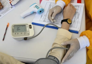
للعاملين في الليل.. حيلة صحية تجنبكم خطر هذا النوع من العمل
|
|
|
|
|
|
|
"ناسا" تحتفي برائد الفضاء السوفياتي يوري غاغارين
|
|
|
|
|
|
|
نحو شراكة وطنية متكاملة.. الأمين العام للعتبة الحسينية يبحث مع وكيل وزارة الخارجية آفاق التعاون المؤسسي
|
|
|