
النبات

مواضيع عامة في علم النبات

الجذور - السيقان - الأوراق

النباتات الوعائية واللاوعائية

البذور (مغطاة البذور - عاريات البذور)

الطحالب

النباتات الطبية


الحيوان

مواضيع عامة في علم الحيوان

علم التشريح

التنوع الإحيائي

البايلوجيا الخلوية


الأحياء المجهرية

البكتيريا

الفطريات

الطفيليات

الفايروسات


علم الأمراض

الاورام

الامراض الوراثية

الامراض المناعية

الامراض المدارية

اضطرابات الدورة الدموية

مواضيع عامة في علم الامراض

الحشرات


التقانة الإحيائية

مواضيع عامة في التقانة الإحيائية


التقنية الحيوية المكروبية

التقنية الحيوية والميكروبات

الفعاليات الحيوية

وراثة الاحياء المجهرية

تصنيف الاحياء المجهرية

الاحياء المجهرية في الطبيعة

أيض الاجهاد

التقنية الحيوية والبيئة

التقنية الحيوية والطب

التقنية الحيوية والزراعة

التقنية الحيوية والصناعة

التقنية الحيوية والطاقة

البحار والطحالب الصغيرة

عزل البروتين

هندسة الجينات


التقنية الحياتية النانوية

مفاهيم التقنية الحيوية النانوية

التراكيب النانوية والمجاهر المستخدمة في رؤيتها

تصنيع وتخليق المواد النانوية

تطبيقات التقنية النانوية والحيوية النانوية

الرقائق والمتحسسات الحيوية

المصفوفات المجهرية وحاسوب الدنا

اللقاحات

البيئة والتلوث


علم الأجنة

اعضاء التكاثر وتشكل الاعراس

الاخصاب

التشطر

العصيبة وتشكل الجسيدات

تشكل اللواحق الجنينية

تكون المعيدة وظهور الطبقات الجنينية

مقدمة لعلم الاجنة


الأحياء الجزيئي

مواضيع عامة في الاحياء الجزيئي


علم وظائف الأعضاء


الغدد

مواضيع عامة في الغدد

الغدد الصم و هرموناتها

الجسم تحت السريري

الغدة النخامية

الغدة الكظرية

الغدة التناسلية

الغدة الدرقية والجار الدرقية

الغدة البنكرياسية

الغدة الصنوبرية

مواضيع عامة في علم وظائف الاعضاء

الخلية الحيوانية

الجهاز العصبي

أعضاء الحس

الجهاز العضلي

السوائل الجسمية

الجهاز الدوري والليمف

الجهاز التنفسي

الجهاز الهضمي

الجهاز البولي


المضادات الميكروبية

مواضيع عامة في المضادات الميكروبية

مضادات البكتيريا

مضادات الفطريات

مضادات الطفيليات

مضادات الفايروسات

علم الخلية

الوراثة

الأحياء العامة

المناعة

التحليلات المرضية

الكيمياء الحيوية

مواضيع متنوعة أخرى

الانزيمات
Hyaline Cartilage
المؤلف:
Neelam Vasudeva and Sabita Mishra
المصدر:
Inderbir Singhs Textbook of Human Histology
الجزء والصفحة:
7th edition , P82-83
2024-12-22
1349
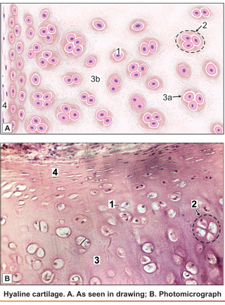
Hyaline cartilage is so called because it is transparent (hyalos = glass).
Its intercellular substance appears to be homogeneous, but using special techniques it can be shown that many collagen fibres are present in the matrix. The fibres are arranged so that they resist tensional forces. Hyaline cartilage has been compared to a tyre. The ground substance (corresponding to the rubber of the tyre) resists compressive forces, while the collagen fibres (corresponding to the treads of the tyre) resist tensional forces.
In haematoxylin and eosin stained preparations, the matrix is stained blue, i.e., it is basophilic. However, the matrix just under the perichondrium is acidophilic .Towards the centre of a mass of hyaline cartilage the chondrocytes are large and are usually present in groups (of two or more). The groups are formed by division of a single parent cell. The cells tend to remain together as the dense matrix prevents their separation. Groups of cartilage cells are called cell-nests (or isogenous cell groups). Towards the periphery of the cartilage, the cells are small and elongated in a direction parallel to the surface. Just under the perichondrium the cells become indistinguishable from fibroblasts.
Immediately around lacunae housing individual chondrocytes, and around cell nests the matrix stains deeper than elsewhere giving the appearance of a capsule. This deep staining matrix is newly formed and is called the territorial matrix or lacunar capsule. In contrast the pale staining matrix separating cell nests is the interstitial matrix.
Calcification of hyaline cartilage is often seen in old people. The costal cartilages or the large cartilages of the larynx are commonly affected. In contrast to hyaline cartilage, elastic cartilage and fibrocartilage do not undergo calcification. Although articular cartilage is a variety of hyaline cartilage, it does not undergo calcification or ossification.
Distribution of Hyaline Cartilage
- Costal cartilages: These are bars of hyaline cartilage that connect the ventral ends of the ribs to the sternum, or to adjoining costal cartilages. They show the typical structure of hyaline cartilage described above. The cellularity of costal cartilage decreases with age.
- Articular cartilage: The articular surfaces of most synovial joints are lined by hyaline cartilage. These articular cartilages provide the bone ends with smooth surfaces between which there is very little friction. They also act as shock absorbers. Articular cartilages are not covered by perichondrium. Their surface is kept moist by synovial fluid that also provides nutrition to them.
Other sites where hyaline cartilage is found:
- The skeletal framework of the larynx is formed by a number of cartilages. Of these the thyroid cartilage, the cricoid cartilage and the arytenoid cartilage are composed of hyaline cartilage.
- The walls of the trachea and large bronchi contain incomplete rings of cartilage. Smaller bronchi have pieces of cartilage of irregular shape in their walls.
- Parts of the nasal septum and the lateral wall of the nose are made up of pieces of hyaline cartilage.
- In growing children long bones consist of a bony diaphysis (corresponding to the shaft) and of one or more bony epiphyses (corresponding to bone ends or projections). Each epiphysis is connected to the diaphysis by a plate of hyaline cartilage called the epiphyseal plate. This plate is essential for bone growth.

 الاكثر قراءة في مواضيع عامة في علم وظائف الاعضاء
 اخر الاخبار
اخبار العتبة العباسية المقدسة

الآخبار الصحية


 قسم الشؤون الفكرية يصدر كتاباً يوثق تاريخ السدانة في العتبة العباسية المقدسة
 "المهمة".. إصدار قصصي يوثّق القصص الفائزة في مسابقة فتوى الدفاع المقدسة للقصة القصيرة
 (نوافذ).. إصدار أدبي يوثق القصص الفائزة في مسابقة الإمام العسكري (عليه السلام)


