![]()


النبات

مواضيع عامة في علم النبات
الجذور - السيقان - الأوراق
النباتات الوعائية واللاوعائية
البذور (مغطاة البذور - عاريات البذور)
الطحالب
النباتات الطبية
الحيوان
مواضيع عامة في علم الحيوان
علم التشريح
التنوع الإحيائي
البايلوجيا الخلوية
الأحياء المجهرية
البكتيريا
الفطريات
الطفيليات
الفايروسات
علم الأمراض
الاورام
الامراض الوراثية
الامراض المناعية
الامراض المدارية
اضطرابات الدورة الدموية
مواضيع عامة في علم الامراض
الحشرات
التقانة الإحيائية
مواضيع عامة في التقانة الإحيائية
التقنية الحيوية المكروبية
التقنية الحيوية والميكروبات
الفعاليات الحيوية
وراثة الاحياء المجهرية
تصنيف الاحياء المجهرية
الاحياء المجهرية في الطبيعة
أيض الاجهاد
التقنية الحيوية والبيئة
التقنية الحيوية والطب
التقنية الحيوية والزراعة
التقنية الحيوية والصناعة
التقنية الحيوية والطاقة
البحار والطحالب الصغيرة
عزل البروتين
هندسة الجينات
التقنية الحياتية النانوية
مفاهيم التقنية الحيوية النانوية
التراكيب النانوية والمجاهر المستخدمة في رؤيتها
تصنيع وتخليق المواد النانوية
تطبيقات التقنية النانوية والحيوية النانوية
الرقائق والمتحسسات الحيوية
المصفوفات المجهرية وحاسوب الدنا
اللقاحات
البيئة والتلوث
علم الأجنة
اعضاء التكاثر وتشكل الاعراس
الاخصاب
التشطر
العصيبة وتشكل الجسيدات
تشكل اللواحق الجنينية
تكون المعيدة وظهور الطبقات الجنينية
مقدمة لعلم الاجنة
الأحياء الجزيئي
مواضيع عامة في الاحياء الجزيئي
علم وظائف الأعضاء
الغدد
مواضيع عامة في الغدد
الغدد الصم و هرموناتها
الجسم تحت السريري
الغدة النخامية
الغدة الكظرية
الغدة التناسلية
الغدة الدرقية والجار الدرقية
الغدة البنكرياسية
الغدة الصنوبرية
مواضيع عامة في علم وظائف الاعضاء
الخلية الحيوانية
الجهاز العصبي
أعضاء الحس
الجهاز العضلي
السوائل الجسمية
الجهاز الدوري والليمف
الجهاز التنفسي
الجهاز الهضمي
الجهاز البولي
المضادات الحيوية
مواضيع عامة في المضادات الحيوية
مضادات البكتيريا
مضادات الفطريات
مضادات الطفيليات
مضادات الفايروسات
علم الخلية
الوراثة
الأحياء العامة
المناعة
التحليلات المرضية
الكيمياء الحيوية
مواضيع متنوعة أخرى
الانزيمات

HeLa Cells
 المؤلف:
D. A. Jackson and A. Pombo
المؤلف:
D. A. Jackson and A. Pombo
 المصدر:
J. Cell Biol. 140, 1285–1295
المصدر:
J. Cell Biol. 140, 1285–1295
 الجزء والصفحة:
 16-5-2016
 3583
3583


HeLa Cells
The HeLa cell line was originated by George Gey and co-workers in 1952 (1) from a cervical adenocarcinoma from a 31-year-old Afro-American female. It has proved to be a very vigorously growing cell line and is one of the most extensively used cell lines in the world. It is lodged at the American Type Culture Collection (ATCC) as CCL-2. It is even more extensively used than many people realize, because it is also guilty of cross-contaminating more cell lines than any other. HeLa was also the first human cell line to be cloned, when HeLa-S3 was isolated by Puck in 1956 (2). HeLa-S3 (Fig. 1) is in the ATCC catalogue as CCL-2.2. HeLa cells were originally propagated in a combination of human and calf serum, but it has been shown that human serum is not necessary, and they are usually maintained in Eagle's Minimal Essential Medium supplemented with 10% fetal bovine serum. HeLa-S3 can be propagated in suspension using low-calcium medium (3).
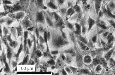
Figure 1. Sub-confluent culture of Hela-S3 cells. Phase contrast, Olympus CK microscope, 20× objective.
1. Properties
HeLa is a continuous cell line with an aneuploid and heteroploid karyotype containing a number of marker chromosomes (4). It expresses the A subtype of glucose-6-phosphate dehydrogenase, which is present in Negroes and absent in Caucasians. The cells are cytokeratin positive, confirming their epithelial origin, contain papilloma virus sequences (HPV-18), and have low expression of p53 and normal retinoblastoma proteins (5). They have a short population doubling time (18–22 h) and high plating efficiency (60–80% in monolayer.(
2. Usage
HeLa cells have been used for propagation and assay of viruses and have been shown to be susceptible to poliovirus-1,2,3, adenovirus-3, encephalomyocarditis, and coxsackievirus B5. They have been used in molecular studies (6, 7), in somatic hybridization analysis of senescence (8) and invasion (9), in fermentor culture for biotechnology (10), and for the development of serum-free media (11). As they are deficient in gap-junction communication, they have been used for transfection analysis of gap-junctional proteins (12). They have also been used for studies on molecular signaling (13. (
HeLa-S3, a clone of HeLa (2), is one of the more rapidly dividing of the HeLa strains and can be grown in suspension with mechanical agitation. Because it is less well attached than the parental strain, mitotic cells are readily detached by shaking the flask, following partial cell-cycle blockade induced by short-term storage at 4°C, (14, 15). H-HeLa is a subline suitable for the passage and titration of rhinoviruses (16).
3. Cross-Contamination
HeLa was the first human cell line to be propagated widely in a large number of laboratories throughout the world. It is also a vigorously growing cell line with a high plating efficiency and is, therefore, ideally placed to cross-contaminate other cell lines. That this happened was first demonstrated many years ago by isoenzyme analysis (17) and subsequently by chromosomal analysis (18). Combined with more recent studies of DNA fingerprinting, it is now clear that a large number of continuous cell lines established in the 1960s and 1970s were cross-contaminated with HeLa cells, including lines such as KB (19) and Hep-2 (19, 20), which are still in regular use. The most serious aspect of this is that, although continued use may be quite acceptable in certain circumstances, few authors acknowledge that these lines are HeLa-contaminated, suggesting that a large part of the scientific community is still unaware of this problem, over 30 years after it was first revealed (21). Reports published by the ATCC (22), and the European Collection of Animal Cell Cultures (ECACC) (23), have confirmed that a significant proportion of cell lines in common use may be HeLa cross-contaminants.
References
1. G. O. Gey, W. D. Coffman, and M. T. Kubicek (1952) Cancer Res. 12, 364–365.
2. T. T. Puck and P. I. Marcus (1955) Proc. Natl. Acad. Sci. USA 41, 432–437.
3. R. I. Freshney (1994) Culture of Animal Cells, a Manual of Basic Technique, Wiley-Liss, New York, p. 85.
4. T. R. Chen (1988) Cytogenet. Cell Genet. 48, 19–24.
5. American Type Culture Collection catalogue, ATCC, P.O. Box 1549, Manassas, VA 20108 –1549 www.atcc.orgñ, CCL-2.
6. W. K. Hansen, W. A. Deutsch, A. Yacoub, Y. Xu, D. A. Williams, and M. R. Kelley (1998) J. Biol. Chem. 273, 756–762.
7. D. A. Jackson and A. Pombo, (1998) J. Cell Biol. 140, 1285–1295.
8. M. D. Waterfield, M. A. Baker, M. F. Greaves, and E. J. Stanbridge (1986) J. Biol. Chem. 261, 2418-2424.
9. K. Ess, H. Chen, A. Kier, and R. Brackenbury (1995) J. Cell. Physiol. 162, 341–347.
10. Y. Chen, T. L. LaPorte, S. S. Wang, and J. Shevitz (1992) Cytotechnology 8, 85–88.
11. G. J. Blaker, J. R. Birch, and S. J. Pirt (1971) J. Cell. Sci. 9, 529–537.
12. B. Hertlein, A. Butterweck, S. Haubrich, K. Willecke, and O. Traub (1998) J. Memb. Biol. 162, 247-257.
13. A. W. Gagnon, L. Kallal, and J. L. Benovic (1998) J. Biol. Chem. 273, 6976–6981.
14. A. A. Newton and P. Wildy (1959) Exp. Cell Res. 16, 624–635.
15. B. Lesser and T. P. Brent (1970) Exp. Cell Res. 62, 470–473.
16. R. R. Rueckert et al. (1971) Virology 44, 259–270.
17. S. M. Gartler (1967) Second Decennial Review Conference on Cell, Tissue and Organ Culture; NCI Monograph, pp. 167–195.
18. W. Nelson-Rees and R. R. Flandermeyer (1977) Science 195, 1343–1344.
19. S. J. O''Brien, R. Olson, and J. E. Shannon (1977) Science 195, 1345–1348.
20. T. R. Chen (1988) Cytogenet. Cell Genet. 48, 19–24.
21. C. S. Stulberg, W. D. Peterson Jr., and W. F. Simpson (1976) Am. J. Hematol. 1, 237–242.
22. K. S. Lavappa (1978) 14, 469–475.
23. G. Stacey, B. Bolton A. Doyle, and B. Griffiths (1992) Cytotechnology 9, 211–216.


 المكياج بلا حدود.. ظاهرة متنامية تُقلق القيم وتُنهك الذات
 جاهزية الاستعداد لشهر رمضان
 إستعراض موجز لحياة السيدة زينب الكبرى


