


النبات

مواضيع عامة في علم النبات
الجذور - السيقان - الأوراق
النباتات الوعائية واللاوعائية
البذور (مغطاة البذور - عاريات البذور)
الطحالب
النباتات الطبية
الحيوان
مواضيع عامة في علم الحيوان
علم التشريح
التنوع الإحيائي
البايلوجيا الخلوية
الأحياء المجهرية
البكتيريا
الفطريات
الطفيليات
الفايروسات
علم الأمراض
الاورام
الامراض الوراثية
الامراض المناعية
الامراض المدارية
اضطرابات الدورة الدموية
مواضيع عامة في علم الامراض
الحشرات
التقانة الإحيائية
مواضيع عامة في التقانة الإحيائية
التقنية الحيوية المكروبية
التقنية الحيوية والميكروبات
الفعاليات الحيوية
وراثة الاحياء المجهرية
تصنيف الاحياء المجهرية
الاحياء المجهرية في الطبيعة
أيض الاجهاد
التقنية الحيوية والبيئة
التقنية الحيوية والطب
التقنية الحيوية والزراعة
التقنية الحيوية والصناعة
التقنية الحيوية والطاقة
البحار والطحالب الصغيرة
عزل البروتين
هندسة الجينات
التقنية الحياتية النانوية
مفاهيم التقنية الحيوية النانوية
التراكيب النانوية والمجاهر المستخدمة في رؤيتها
تصنيع وتخليق المواد النانوية
تطبيقات التقنية النانوية والحيوية النانوية
الرقائق والمتحسسات الحيوية
المصفوفات المجهرية وحاسوب الدنا
اللقاحات
البيئة والتلوث
علم الأجنة
اعضاء التكاثر وتشكل الاعراس
الاخصاب
التشطر
العصيبة وتشكل الجسيدات
تشكل اللواحق الجنينية
تكون المعيدة وظهور الطبقات الجنينية
مقدمة لعلم الاجنة
الأحياء الجزيئي
مواضيع عامة في الاحياء الجزيئي
علم وظائف الأعضاء
الغدد
مواضيع عامة في الغدد
الغدد الصم و هرموناتها
الجسم تحت السريري
الغدة النخامية
الغدة الكظرية
الغدة التناسلية
الغدة الدرقية والجار الدرقية
الغدة البنكرياسية
الغدة الصنوبرية
مواضيع عامة في علم وظائف الاعضاء
الخلية الحيوانية
الجهاز العصبي
أعضاء الحس
الجهاز العضلي
السوائل الجسمية
الجهاز الدوري والليمف
الجهاز التنفسي
الجهاز الهضمي
الجهاز البولي
المضادات الميكروبية
مواضيع عامة في المضادات الميكروبية
مضادات البكتيريا
مضادات الفطريات
مضادات الطفيليات
مضادات الفايروسات
علم الخلية
الوراثة
الأحياء العامة
المناعة
التحليلات المرضية
الكيمياء الحيوية
مواضيع متنوعة أخرى
الانزيمات

PHYSIOLOGIC ANATOMY OF CARDIAC MUSCLE
 المؤلف:
John E. Hall, PhD
المؤلف:
John E. Hall, PhD
 المصدر:
Guyton and Hall Textbook of Medical Physiology
المصدر:
Guyton and Hall Textbook of Medical Physiology
 الجزء والصفحة:
13th Edition , p109-110
الجزء والصفحة:
13th Edition , p109-110
 2025-02-04
 1043
1043


+
-
20
Figure 1 shows the histology of cardiac muscle, demonstrating cardiac muscle fibers arranged in a latticework, with the fibers dividing, recombining, and then spreading again. Note that cardiac muscle is striated in the same manner as in skeletal muscle. Further, cardiac muscle has typical myofibrils that contain actin and myosin filaments almost identical to those found in skeletal muscle; these filaments lie side by side and slide during contraction in the same manner as occurs in skeletal muscle . In other ways, however, cardiac muscle is quite different from skeletal muscle, as we shall see.
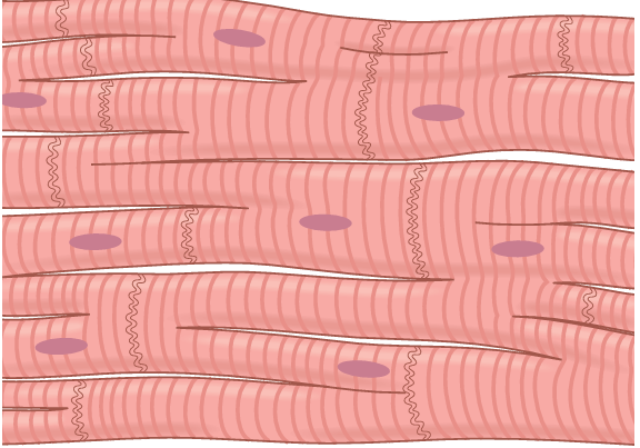
fig1. Syncytial, interconnecting nature of cardiac muscle fibers.
Cardiac Muscle Is a Syncytium. The dark areas crossing the cardiac muscle fibers in Figure 1 are called intercalated discs; they are actually cell membranes that separate individual cardiac muscle cells from one another. That is, cardiac muscle fibers are made up of many individual cells connected in series and in parallel with one another.
At each intercalated disc the cell membranes fuse with one another to form permeable “communicating” junctions (gap junctions) that allow rapid diffusion of ions. Therefore, from a functional point of view, ions move with ease in the intracellular fluid along the longitudinal axes of the cardiac muscle fibers so that action potentials travel easily from one cardiac muscle cell to the next, past the intercalated discs. Thus, cardiac muscle is a syncytium of many heart muscle cells in which the cardiac cells are so interconnected that when one cell becomes excited, the action potential rapidly spreads to all of them.
The heart actually is composed of two syncytiums: the atrial syncytium, which constitutes the walls of the two atria, and the ventricular syncytium, which constitutes the walls of the two ventricles. The atria are separated from the ventricles by fibrous tissue that surrounds the atrioventricular (A-V) valvular openings between the atria and ventricles. Normally, potentials are not con ducted from the atrial syncytium into the ventricular syncytium directly through this fibrous tissue. Instead, they are conducted only by way of a specialized conductive system called the A-V bundle, a bundle of conductive fibers several millimeters in diameter.
This division of the muscle of the heart into two functional syncytiums allows the atria to contract a short time ahead of ventricular contraction, which is important for effectiveness of heart pumping.
 الاكثر قراءة في الجهاز الدوري والليمف
 اخر الاخبار
اخبار العتبة العباسية المقدسة

الآخبار الصحية


مواضيع ذات صلة
 قسم الشؤون الفكرية يصدر كتاباً يوثق تاريخ السدانة في العتبة العباسية المقدسة
 "المهمة".. إصدار قصصي يوثّق القصص الفائزة في مسابقة فتوى الدفاع المقدسة للقصة القصيرة
 (نوافذ).. إصدار أدبي يوثق القصص الفائزة في مسابقة الإمام العسكري (عليه السلام)


